
Heart anatomy in 3D
BetaTurn the heart in any direction, label its chambers, valves and vessels, cut through it in three planes and follow blood step by step through a 12-step tour.
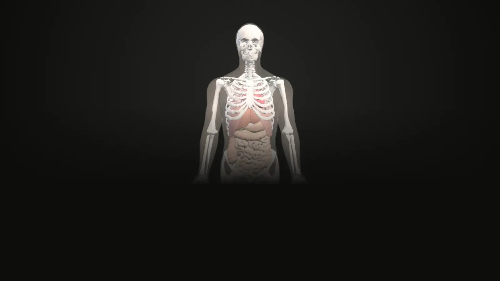
Interactive 3D atlas
Loads about 9 MB of 3D data the first time. Works best on a laptop or a recent phone.
Anatomy Atlas
Layers
Heart
Organ workstationColour parts by
Section cut
Elevation 0°
In the chest
Where everything in this atlas comes from
Study the heart in 3D
Turn the heart in any direction, label its chambers, valves and vessels, cut through it in three planes and follow blood step by step through a 12-step tour.
Turn and zoom
Drag to rotate, scroll or pinch to zoom, and tap any part to see its name and explanation.
Labels and standard views
Switch labels on or off and jump to front, back, side, top and bottom views, with an orientation cube.
Section cuts
Cut through the structure in transverse, coronal or sagittal planes and slide the cut through it.
Explode and see-through
Pull the parts apart, or make the outer layers faint to see what is inside.
Guided tour
Follow the "Blood flow" tour one step at a time, or press Play and let the camera fly to each part.
Parts in this model
99 named parts, each with the FMA identifier the dataset gives it. Tap a group in the app to show or hide it.
- Chamber walls3
- Chamber cavities4
- Valve leaflets and cusps11
- Papillary muscles5
- Coronary arteries54
- Cardiac veins7
- Great arteries6
- Great veins9
Guided tour: Blood flow (12 steps)
Each step highlights the parts it talks about. The sentence is quoted word for word from OpenStax Anatomy and Physiology 2e.
1. Superior vena cava, Inferior vena cava
The blood in the superior and inferior venae cavae flows into the right atrium, which pumps blood into the right ventricle.
OpenStax, 19.1 Chambers and Circulation through the Heart2. Cavity of right atrium
The opening between the atrium and ventricle is guarded by the tricuspid valve.
OpenStax, 19.1 Right Atrium3. Anterior leaflet of tricuspid valve, Posterior leaflet of tricuspid valve, Septal leaflet of tricuspid valve
The right ventricle receives blood from the right atrium through the tricuspid valve.
OpenStax, 19.1 Right Ventricle4. Cavity of right ventricle
The right ventricle pumps deoxygenated blood into the pulmonary trunk, which leads toward the lungs and bifurcates into the left and right pulmonary arteries.
OpenStax, 19.1 Chambers and Circulation through the Heart5. Left anterior cusp of pulmonary valve, Right anterior cusp of pulmonary valve, Posterior cusp of pulmonary valve
At the base of the pulmonary trunk is the pulmonary semilunar valve that prevents backflow from the pulmonary trunk.
OpenStax, 19.1 Right Ventricle6. Pulmonary trunk, Left pulmonary artery, Right pulmonary artery
When the right ventricle contracts, it ejects blood into the pulmonary trunk, which branches into the left and right pulmonary arteries that carry it to each lung.
OpenStax, 19.1 Right Ventricle7. Left inferior pulmonary vein, Left superior pulmonary vein, Right inferior pulmonary vein
After exchange of gases in the pulmonary capillaries, blood returns to the left atrium high in oxygen via one of the four pulmonary veins.
OpenStax, 19.1 Left Atrium8. Cavity of left atrium
The opening between the left atrium and ventricle is guarded by the mitral valve.
OpenStax, 19.1 Left Atrium9. Anterior leaflet of mitral valve, Posterior leaflet of mitral valve
Located at the opening between the left atrium and left ventricle is the mitral valve, also called the bicuspid valve or the left atrioventricular valve.
OpenStax, 19.1 Heart Valve Structure and Function10. Cavity of left ventricle
The left ventricle is the major pumping chamber for the systemic circuit; it ejects blood into the aorta through the aortic semilunar valve.
OpenStax, 19.1 Left Ventricle11. Anterior cusp of aortic valve, Left posterior cusp of aortic valve, Right posterior cusp of aortic valve
At the base of the aorta is the aortic semilunar valve, or the aortic valve, which prevents backflow from the aorta.
OpenStax, 19.1 Heart Valve Structure and Function12. Ascending aorta, Arch of aorta, Descending thoracic aorta
The pulmonary veins conduct blood into the left atrium, which pumps the blood into the left ventricle, which in turn pumps oxygenated blood into the aorta and on to the many branches of the systemic circuit.
OpenStax, 19.1 Chambers and Circulation through the Heart
What this model does not show
The great vessels are cut short near the heart, and some small heart vessels have no passage of their own in the textbook, so the page shows the passage of their group.
Where the data or the textbook has a gap, the atlas says so instead of filling it in.
Keep exploring
You moved to another screen of the atlas. Read the study notes for it on its own page: the atlas.
Questions
What can I see in the heart model?
The model has 99 named parts in 8 groups: Chamber walls, Chamber cavities, Valve leaflets and cusps, Papillary muscles, Coronary arteries, Cardiac veins, Great arteries, Great veins.
What is the "Blood flow" tour?
It is a guided tour of 12 steps. Each step highlights the parts it describes and shows a sentence quoted from OpenStax Anatomy and Physiology 2e. Press Play to let the camera fly to each step for you.
What is missing from the heart model?
The great vessels are cut short near the heart, and some small heart vessels have no passage of their own in the textbook, so the page shows the passage of their group.
Where does the information come from?
The 3D shapes come from the BodyParts3D dataset (and, for the female pelvic region, the Human Reference Atlas). Every explanation is a sentence quoted word for word from the open textbook OpenStax Anatomy and Physiology 2e, with a link to its section.
Can I use it to diagnose or treat anything?
No. The atlas is for learning only. It is a beta version and has not yet been reviewed by an anatomy educator.
Credits and disclaimer
For learning only, not for diagnosis or treatment. This is a beta version and it has not yet been reviewed by an anatomy educator.
BodyParts3D, © The Database Center for Life Science licensed under CC Attribution 4.0 International. Text quoted from OpenStax, Anatomy and Physiology 2e, © Rice University, CC BY-NC-SA 4.0. Female pelvic region from the Human Reference Atlas (HuBMAP), CC BY 4.0.
This tool is free, non-commercial and made for education. It has no ads and no paid features.